
What are Glycosaminoglycans?
GAGS Glycosaminoglycans are the hetero-polysaccharides of Extra Cellular Matrix (ECM) of Cells. ECM is filled with a gel-like material and it is the ground substance which holds adjacent cells. ECM gives a porous pathway for nutrients and oxygen. Glycosaminoglycans are often denoted as GAGs. Glycosaminoglycans are unique to animals and bacteria and are NOT found in plants. The present post discusses the structure, characteristics, classification and functions of Glycosaminoglycans with suitable examples.
Table of Contents
@. Structure of Glycosaminoglycans
@. Examples of GAGS
$. Hyaluronic Acid (Hyaluronan)
$. Chondroitin sulfate
$. Dermatan sulfate
$. Keratan sulfate
$. Heparan sulfate
@. Related Posts
Structure of Glycosaminoglycans (GAGs)
Ø Glycosaminoglycans are linear hetero-polysaccharides of ECM.
Ø They composed of repeating disaccharide units.
Ø One of the monosaccharides in the disaccharide unit is always either:
$. N-acetyl glucosamine
$. N-acetylgalactosamine
Ø The other monosaccharide residue in most cases will be a uronic acid usually a D-glucuronic acid or L-iduronic acid (Learn more: Sugar Derivatives).
Ø In some glycosaminoglycans, one or more hydroxyl groups of the amino sugars are esterified with sulphate.
Ø The sulphate group gives a negative charge to the molecule.
| You may also like NOTES in... | ||
|---|---|---|
| BOTANY | BIOCHEMISTRY | MOL. BIOLOGY |
| ZOOLOGY | MICROBIOLOGY | BIOSTATISTICS |
| ECOLOGY | IMMUNOLOGY | BIOTECHNOLOGY |
| GENETICS | EMBRYOLOGY | PHYSIOLOGY |
| EVOLUTION | BIOPHYSICS | BIOINFORMATICS |
Ø The combination of sulfate groups and the carboxylate groups of the uronic acid gives glycosaminoglycans a very high density of negative charges.
Ø To minimize the repulsive forces among neighboring charged groups, the glycosaminoglycan molecules assume an extended conformation in solution.
Ø This extended conformation usually forms a rod-like helix in which the negatively charged carboxylate groups occur on alternate sides of the helix.
Ø The extended rod form also provides maximum separation between the negatively charged sulfate groups.
Ø The specific patterns of sulfated and non-sulfated sugar residues in glycosaminoglycans provide specific recognition by a variety of protein ligands.
Ø These protein ligands bind electrostatically to the glycosaminoglycan molecules.
Ø The sulfated glycosaminoglycans are attached to extracellular proteins to form proteoglycans (a glycoconjugate).
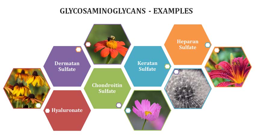
Examples of glycosaminoglycans:
(1). Hyaluronate (Hyaluronan)
(2). Chondroitin sulfate
(a). Chondroitin-4-sulphate
(b)· Chondroitin-6-sulphate
(3). Dermatan sulfate
(4). Keratan sulfate
(5). Heparan sulfate
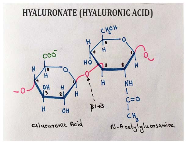
(1). Hyaluronic Acid
Ø They are also called as hyaluronate.
Ø Hyaluronate is the most abundant glycosaminoglycan in the cells.
Ø It is a non-sulfated anionic glycosaminoglycan.
Ø It consists of alternating disaccharide units of D-glucuronic acid and N-acetylglucosamine (50,000 repeats).
Ø The glycosidic bonds that connect these monomeric units are:
$. β(1→3) : Between glucuronic acid and NAG (disaccharide)
$. β(1→4) : Between disaccharide units
Ø Hyaluronate form a clear highly viscous solution.
Ø They serve as the vitreous humor of eye.
Ø They have high shock absorbing capacity, hence; form the lubricants in synovial fluids (joints).
Ø They are the essential components of cartilage.
Ø Many bacteria produce hyaluronidase enzyme (to enter into the host cell).
Ø Human sperm cells produce hyaluronidase to hydrolyze the thick glycosaminoglycan coat of ovum (Zona pellucida).
Ø Other glycosaminoglycans differ from hyaluronan in FOUR aspects:
(1). They are generally much shorter polymers.
(2). They are covalently linked to specific proteins (proteoglycans).
(3). One or both monomeric units differ from those of hyaluronan.
(4). Have one or more sulfate groups (hence having a negative charge).
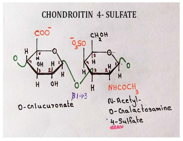
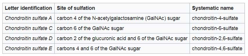
(2). Chondroitin sulfate
Ø Chondroitin sulfate is a sulfated glycosaminoglycan.
Ø It composed of alternating N-acetylgalactosamine and glucuronic acid.
Ø Usually, chondroitin sulfate is found attached to proteins as part of a proteoglycan.
Ø They have over 100 individual sugars, each of which can be sulfated in variable positions and quantities.
Ø Chondroitin sulfate is an important structural component of cartilage.
Ø In the cartilage, chondroitin sulfate provides resistance to compression.
Ø It is also responsible for the high tensile strength of cartilage, tendon, ligaments and walls of the aorta.
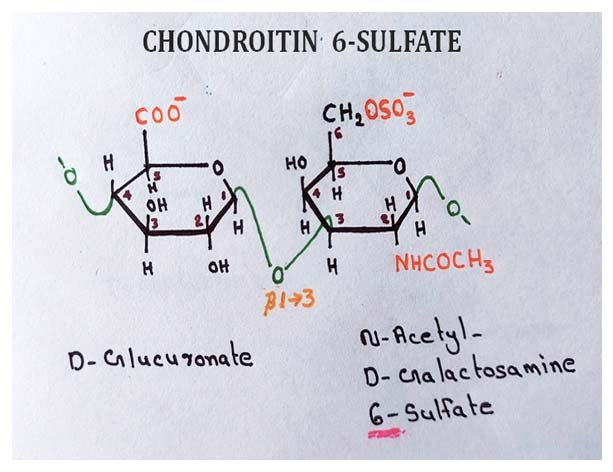
Ø There are different types of chondroitin sulfate in the cell based on the position of sulfate group(s).
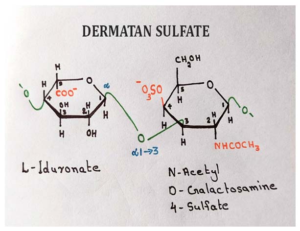
(3). Dermatan Sulfate
Ø Dermatan sulfate is also called as chondroitin sulfate B.
Ø It is mostly found in the skin.
| You may also like... | ||
|---|---|---|
| NOTES | QUESTION BANK | COMPETITIVE EXAMS. |
| PPTs | UNIVERSITY EXAMS | DIFFERENCE BETWEEN.. |
| MCQs | PLUS ONE BIOLOGY | NEWS & JOBS |
| MOCK TESTS | PLUS TWO BIOLOGY | PRACTICAL |
Ø Dermatan sulfate is also in blood vessels, heart valves and lungs.
Ø Dermatan sulfate is derived from chondroitin by enzymatic epimerization of the C5 of glucuronate residues to form iduronate residues.
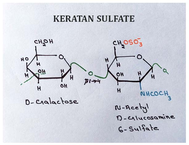
(4). Keratan sulfates
Ø Ketatan sulfates should NOT be confused with the fibrous protein keratin.
Ø Keratan sulfate is the most heterogeneous glycosaminoglycan.
Ø Its sulfate content is highly variable and it contains small amounts of fucose, mannose, N-acetyl glucosamine, and sialic acid. (hence they includes many classes)
Ø Keratan sulfate usually found in cornea, cartilage and bones
Ø It has very good shock absorbing capacity.
(5). Heparan sulfate
Ø Heparan sulfate is a complex glycosaminoglycan produced by all animal cells and contains variable arrangements of sulfated and non-sulfated sugars.
Ø The sulfated segments of the chain allow it to interact with a large number of proteins, including growth factors and ECM components.
Ø Heparin is a fractionated form of heparan sulfate derived mostly from mast cells.
Ø Mast cells are a type of leukocyte.
Ø Heparin is a therapeutic agent used to inhibit blood coagulation through its capacity to bind the protease inhibitor anti-thrombin.
Ø Heparin binding causes anti-thrombin to bind to and inhibit thrombin, a protease essential to blood clotting.
Ø This interaction is strongly electrostatic.
Ø Heparin has the highest negative charge density of any known biological macromolecule.
Ø Purified heparin is routinely added to blood samples obtained for clinical analysis, and to blood donated for transfusion, to prevent clotting.
References
@. Lehninger A.B., (2018), Textbook of Biochemistry, Ed. 5, Pearson International, New York
@. Voet, D., Voet, J.G. and Pratt, C.W., 2013. Fundamentals of biochemistry: life at the molecular level
Do you have any Queries? Please leave me in the Comments Section below.
I will be Happy to Read your Comments and Reply.
<<< Back to Biochemistry Lecture Notes Home Page
Introduction to Carbohydrates | Monosaccharides | Disaccharides | Polysaccharides | Glycosaminoglycans | Sugar Derivatives | Glycoconjugates |
More Lecture Notes from Easy Biology Class…
BotanyZoologyBiochemistryGeneticsCell & Molecular BiologyBiotechnologyPhysiology & EndocrinologyPlant PhysiologyMicrobiologyImmunologyEmbryologyEcologyEvolutionBiophysicsResearch MethodologyBiostatisticsChemistry for BiologistsPhysics for Biologists
Browse more in Easy Biology Class…
Lecture NotesBiology PPTsVideo TutorialsBiology MCQQuestion BankDifference betweenPractical AidsMock Tests (Online)Biology Exams
This notese is better than our biochemistry teacher
Hi, how can I access protected content,?
The notes are very helpful.