
Flowering plants (Angiosperms) show sexual reproduction. Flowers are the important reproductive structure produced by Angiosperms to affect sexual reproduction. Flowers are the objects of aesthetic and ornamental value, but for a botanist, flowers are the sites of sexual reproduction. This post is the Part-1 of Plus Two Botany Notes Sexual Reproduction in Flowering Plants. Here we briefly discuss the parts of a typical flower and Pre-fertilization structures and events.
FLOWER
- Flowers are the reproductive organs in angiosperms or Flowering Plants.
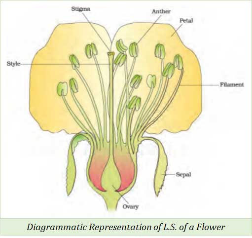
STRUCTURE OF A FLOWER
- A flower has 4 whorls.
- These are calyx, corolla, androecium and gynoecium arranged from outside to inside.
- Components of floral whorls:
- Calyx – Composed of Sepals
- Corolla – Composed of Petals
- Androecium – Composed of Stamens
- Gynoecium – Composed of Carpels / Pistils
- Among these four whorls, calyx and corolla are non-reproductive whorls (non-essential whorls) and Androecium and Gynoecium are reproductive whorls (essential whorls).
REPRODUCTIVE STRUCTURES OR REPRODUCTIVE WHORLS IN A FLOWER
- Usually, a flower has two reproductive whorls.
-
- Male Reproductive whorl: Androecium
- Female Reproductive whorl: Gynoecium
(A). ANDROECIUM
- Androecium is the male reproductive structure of a flower
- Androecium composed of stamens
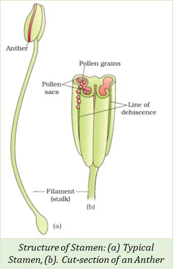
Structure of Stamen
- Each stamen consists of Three parts – Filaments, Anther and Connective
Filament (Stalk)
- The long slender stalk of the stamen
- The filament holds the anther at the tip
Anther
- Anther is a bilobed (two lobed) structure attached to the filament at the tip.
- Each anther has 4 microsporangia (2 per lobe).
- Microsporangia contain pollen grains or microspores.
- Microsporangia develop further and become pollen sacs.
Connective
- Connective is a sterile tissue that connects the two anther lobes.
Structure of Microsporangium
- Microsporangium has four wall layers and a central mass of Sporogenous cells
- The four wall layers of microsporangium are:
-
- Epidermis
- Endothecium
- Middle layers
- Tapetum
Epidermis
-
- Epidermis is the outermost wall layer of microsporangium.
- It is single layered structure
- Function: Protection of microsporangium
Endothecium
- Endothecium is the second wall layer arranged internal to epidermis.
- Function: Protection of microsporangium and dehiscence of anther.
Middle layers
- It is the third layer of microsporangium
- Middle layer consists of 1-3 layers of cells
- Function: Protection of microsporangium and dehiscence
Tapetum
- It is the innermost wall layer of microsporangium
- Cells of the tapetum are multinucleated and they have dense cytoplasm
- Function: Provides nourishment to the developing pollen grains.
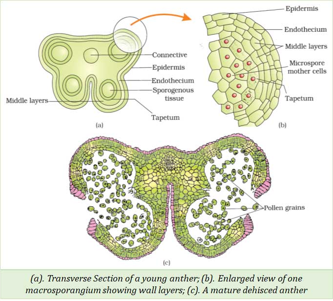
Sporogenous Cells
- The centre of each microsporangium is filled with a closely arranged similar cells called sporogenous cells.
- Sporogenous cells are diploid, pollen grains are formed from the sporogenous cells.
- At maturity, pollen sac is formed by the fusion of two microsporangium in each lobe.
Microsporogenesis
- Microsporogenesis is the process of formation of microspores or pollen grains.
- Microspores are formed from Pollen Mother Cell (PMC) by meiosis (reduction division).
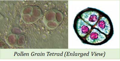 Steps of Microsporogenesis
- Each cell of the Sporogenous tissue (diploid) in a microsporangium acts as Pollen Mother Cell (PMC) or Microspore Mother Cell (2n).
- Pollen Mother Cell (Diploid, 2n) undergoes meiotic cell division to form four haploid (n) cells.
- These four cells are clustered (attached to each other) and hence they are called Microspore Tetrad.
- The anther dehydrates (lost water content) on attaining maturity.
- The microspores in the tetrad get separated from each other to form haploid Pollen Grains (n).
Microsporogenesis: Terms in the Order of Developmental Sequence
- Anther → Sporogenous Tissue → Pollen Mother Cell → Microspore Tetrad → Microspores (Pollen grains) → Male gamete
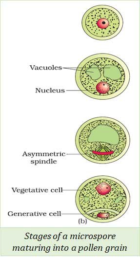 Developmental Stages of a Microspore Maturing into a Pollen Grain
- A newly formed microspore has dense cytoplasm and a central nucleus.
- The central nucleus is pushed towards the periphery by the development of vacuoles (air space).
- The protoplast divides mitotically and form 2 unequal cells
- A bigger cell – Vegetative Cell
- A smaller cell – Generative Cell
- This stage of the pollen grain is called two celled stage.
- In most of the angiosperms, pollen grains are released at this stage.
- But in some angiosperm species, they are released at three celled stages.
- In this case, the generative cell again divides.
Pollen Grains
- Pollen grains develop from the Pollen Mother Cell (2n)
- They represent the Male Gametophyte
- Because they carry the male gametes.
- They are haploid (n)
- Because they are formed by meiotic cell division
Structure of Pollen Grain
- Pollen grains are spherical in shape
- It has two wall layers: (1). Exine and (2). Intine
Exine: The outer wall layer
- Exine is a hard layer
- It is made up of
- Sporopollenin is the most resistant organic material present in nature.
- Because of the tough nature of sporopollenin, pollen grains will be well preserved even in fossils.
- Exine has an aperture called germ pore.
- Sporopollenin is absent at the region of germ pore.
Intine: The inner wall layer
- Intine is made up of Cellulose and Pectin
- Intine is thin layer.
- Each pollen has 2 unequal cells inside (Two Celled stage of Pollen Grain)
- Vegetative Cell
- Vegetative cell is larger in size.
- It is rich in reserve food materials.
- Generative Cell
- Generative cell is smaller in size
- It is spindle shaped.
- It contains dense cytoplasm with a prominent nucleus.
Pollen Viability
- Pollen viability is the time period for which the pollen grains remain functional.
- Examples:
- Rice, Wheat: Pollen viability is 30 minutes (i.e., after 30 minutes the pollen grains of rice and wheat will be non-viable or dead)
- Rosaceae, Leguminosae and Solanaceae plants: viability can be for some months.
Pollen Bank
- Pollen bank is a method to store pollen grains for future use.
- They can be stored in liquid nitrogen (-1960C) for years (Cryopreservation)
- Stored pollen can be used for future plant breeding programmes.
Pollen Allergy
- Pollen grains of some plants cause allergy and respiratory disorders like asthma and bronchitis.
- Example: Parthenium or Carrot grass
Pollen Products
- Pollen grains are rich source of nutrients and unsaturated fats.
- So, they are used to make food supplements in the form of tablets and syrups.
- It increases the performance of athletes and race horse.

(B). GYNOECIUM
- Gynoecium is the female reproductive whorl of the flower
- It is composed of carpel or pistil.
Structure of Pistil or Carpel
- Each pistil has three distinct parts.
Stigma
- Stigma receives pollen grains during pollination.
- The deposited pollen grains get attached to the stigma due to its sticky nature.
Style
- Style is the connecting stalk between stigma and ovary.
Ovary
- Ovary is the basal portion of the carpel.
- It is swollen in shape and contains ovules (megasporangia) attached to the placenta.
- Number of pistils or carpels in the gynoecium
- Gynoecium with single carpel: Monocarpellary
- Gynoecium with many carpels: Multicarpellary
- Multicarpellary gynoecium with fused carpels: Syncarpous
- Multicarpellary gynoecium with free carpels: Apocarpous
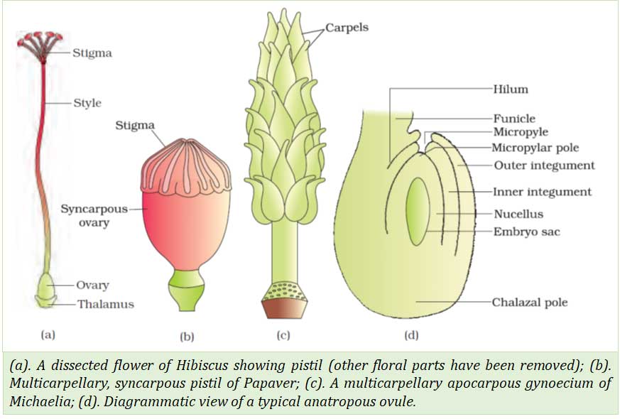
Structure of Ovary
- Ovary is the basal swollen part of the pistil.
- Ovary contains ovules (megasporangium).
- Ovules are attached to the placenta.
Structure of Megasporangium (Ovule)
- The ovule or megasporangium consists of :-
(1). Funicle
- Funicle is the stalk of the ovule.
- It is attached to the placenta on ovary.
(2). Hilum
- m is the junction between the funicle and ovule.
(3). Integuments
- Integuments are the protective envelops around the ovule.
- Two layers of integuments are Outer integument and Inner integument
(4). Micropylar pole
- It is the small opening at the tip of the ovule.
- At this point, integuments do not cover the ovule.
(5). Nucellus
- Nucellus is the layer of cells inside the integuments.
- It is rich in reserve food materials.
(6). Embryosac
- Embryosac is an oval structure within the nucellus.
- It is the female gametophyte of the plant.
- It carries the female gamete or the Egg.
(7). Chalazal pole
- Chalazal pole is th basal part of the ovule.
- It lies opposite to the micropylar pole.
MEGASPOROGENESIS
- Megasporogenesis is the process of formation of haploid megaspores from diploid Megaspore Mother Cell (MMC).
Steps of Megasporogenesis
- A single cell of the nucellus at the micropylar end is differentiated into Megaspore Mother Cell (MMC) or Megasporocyte (2n).
- Megaspore Mother Cell (2n) undergoes meoitic cell division and produces four haploid (n) megaspores.
- Four megaspores are arranged in a linear tetrad
- Among these four megaspores, three lying towards the micropylar end degenerate
- The remaining one megaspore becomes the functional megaspore
- The functional megaspore differentiates into the Embryo sac or the female gametophyte (This type of embryo sac development is called Monosporic development)
- Development of Female Gametophyte or Embryo sac in angiosperms is called Monosporic development. Because:
- It develops from the single functional megaspore (n)
- Other three megaspores towards the micropylar end get degenerated.
DEVELOPMENT OF FEMALE GAMETOPHYTE OR EMBRYOSAC
- The embryo sac develops from the functional megaspore.
- The functional megaspore enlarges in size and its haploid nucleus undergoes three repeated mitotic divisions.
- Thus, eight haploid nuclei are formed.
- These eight nuclei are arranged in such an order to form the following:
- Antipodal cells (3 nuclei)
- Egg apparatus (1 Egg + 2 Synergids)
- Polar nuclei (2 nuclei)
- Antipodal Cells
- Three nuclei move towards the chalazal end of the ovule to form antipodal cells.
- Egg Apparatus
- Three nuclei move towards the micropylar end to form the Egg Apparatus.
- The egg apparatus consists of 2 synergids and 1
- The central nucleus develops into egg.
- The two nuclei on the sides of the egg develops into synergids or
- Function of Synergids: To direct the pollen tube into the embryo sac.
- Filiform apparatus: The special cellular thickenings found at the micropylar end of the synergids is called filiform apparatus.
- Function of filiform apparatus: To direct or guide the pollen tube into the synergid.
- Polar Nuclei
- Two nuclei move towards the centre become polar nuclei.
- These two haploid nuclei fuse together to form the diploid secondary fusion nucleus (2n).
- Cell walls are formed around these nuclei except the polar nuclei.
- Thus, a mature embryosac is 7 celled and 8 nucleate.
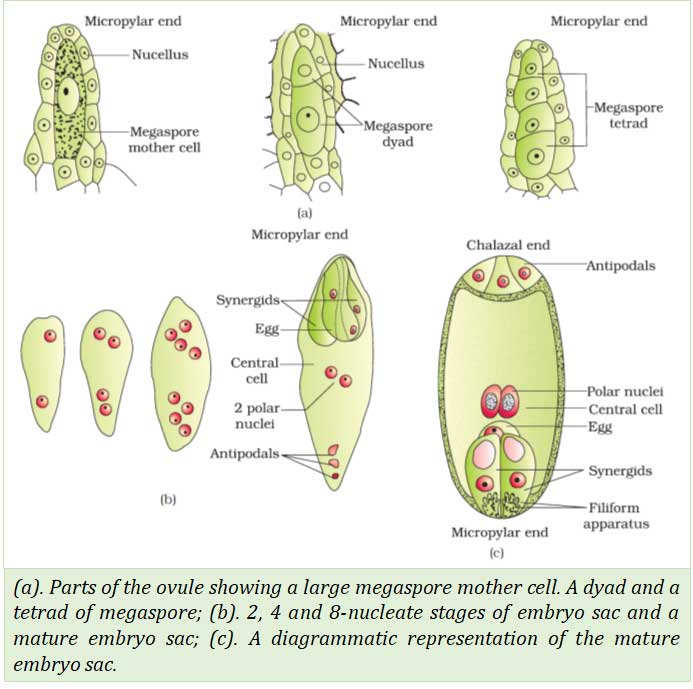
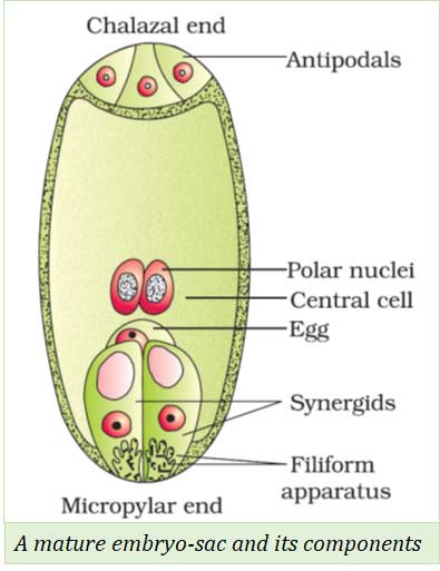
Structure of Embryo sac
- A mature embryosac has seven cells and eight nuclei
- 3 antipodal cells – at the chalazal end
- 1 central cell (with 2 polar nuclei)
- Egg apparatus with 2 synergids and 1 egg – at the micropylar end
- Synergids have some special cellular thickenings called filiform apparatus.
 So far, we have discussed the Flower and Pre-reproductive structure of the flower. In the next post we will see the Pollination, Fertilization and Double Fertilization. You can Download the PDF of this note from the link given below. Hope this post was helpful for your learning. We would like to hear it from you. Please COMMENT (below ↓)
<<< Back to PLUS TWO BOTANY NOTES Page
Study Offline (Without Internet): Now you can download the PDF of this post absolutely free, please follow this link. Please Share the PDF with your Friends.
You might also like…
@. Plus Two Biology Previous Year Question Papers
@. Plus One Biology Previous Year Question Papers
@. Model Question Papers
@. NEET Previous Year Solved Question Papers