
Nipah virus (NiV) is a zoonotic pathogen that has caused outbreaks in several Asian countries. Nipah belongs to the genus Henipavirus. The virus was named after the Malaysian village where it was first identified in 1999. With its high mortality rate and potential for human-to-human transmission, Nipah virus poses a significant threat to public health. Due to the high fatality, transmission, and unavailability of effective treatment and vaccine against the virus, it is considered a Biosafety level 4 (BSL 4) pathogen. In this article ‘Structure of Nipah Virus’ we will explore the ultrastructure, genome, and mechanism of infection of Nipah virus.
Microbiology Notes | Microbiology PPTs | Microbiology MCQs
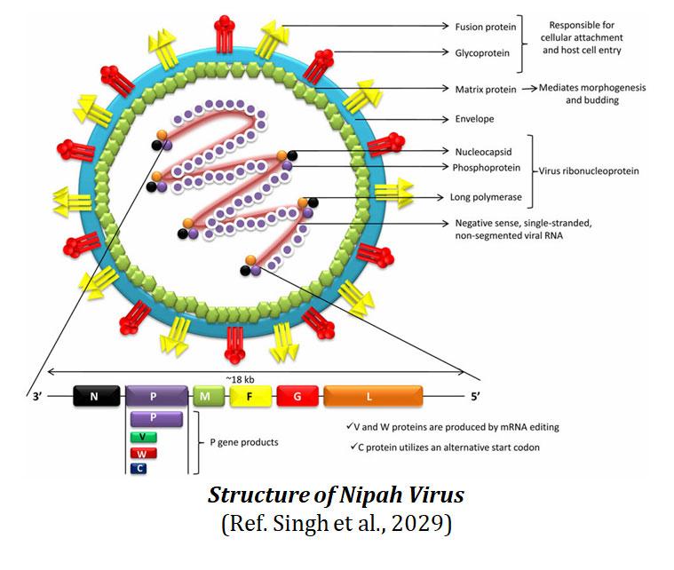
Structure of Nipah Virus
Ø The Nipah virus falls within the Paramyxoviridae family which is characterized by the presence of viral envelope.
Ø Enveloped virus particles are variable in shape and they can be filamentous or spherical.
Ø It possesses a non-segmented, single-stranded RNA genome with a negative-sense, arranged in a helical nucleocapsid structure.
You may also like: Nipah Disease- Transmission, Symptoms, Diagnostics and Treatment
Ø This viral ribonucleoprotein is composed of the nucleocapsid, phosphoprotein, and a long polymerase.
Ø The nucleocapsid is enclosed by a matrix protein housing fusion proteins and glycoproteins that extend outward as spikes
Ø The spikes play a crucial role in attaching to and entering host cells.
Ø In terms of size, Nipah viruses are generally larger than typical paramyxoviruses, ranging from 40 to 1900 nanometers.
Ø Moreover, Nipah virus stands apart from other paramyxoviruses due to the presence of reticular cytoplasmic inclusions near the endoplasmic reticulum.
Ø Nipah virus bears a notable resemblance to the Hendra virus (HeV), displaying only minor differences in ultrastructure
Ø There are two distinct strains of Nipah virus, the Malaysian (MY) and Bangladesh (BD) strains
Ø Both these strains share around 92% sequence similarity but exhibiting variations in their pathogenicity and transmission characteristics.
You may also like NOTES in...
BOTANY BIOCHEMISTRY MOL. BIOLOGY
ZOOLOGY MICROBIOLOGY BIOSTATISTICS
ECOLOGY IMMUNOLOGY BIOTECHNOLOGY
GENETICS EMBRYOLOGY PHYSIOLOGY
EVOLUTION BIOPHYSICS BIOINFORMATICS
Genome of Nipah Virus
Ø Nipah is an enveloped RNA virus.
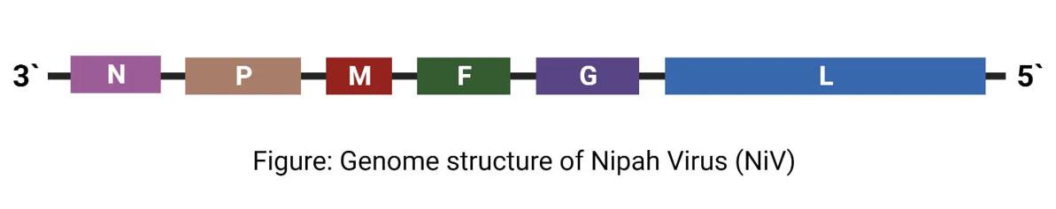
Ø The genome is a single (non-segmented) negative-sense, single-stranded RNA of 18.2 kb size.
Ø Six structural proteins are generated by the genome, namely N (nucleocapsid), P (phosphoprotein), M (matrix), F (fusion), G (glycoprotein) and L (RNA polymerase).
Ø The P open reading frame also encodes three non-structural proteins, V, W and C.
Ø V and W are coded by by RNA editing where as C is coded by an alternate open reading frame.
Ø There are two envelope glycoproteins namely, G glycoprotein and F glycoprotein.
Ø G glycoprotein ectodomain assembles as a homo-tetramer to form the viral anti-receptor or attachment protein, which binds to the receptor on the host cell.
Ø F glycoproteins forms a trimer, which mediates membrane fusion.
Ø The G protein head domain is highly antigenic, inducing specific antibodies in primate models.
Ø G and F proteins are responsible for cellular attachment and entry into the host cells.
Ø The G protein is a prime target for vaccine development as well as antibody therapy.
Ø Ephrins B2 and B3 have been identified as the main receptors for Nipah virus in human.
Replication of Nipah Virus (NiV)
Attachment and Adsorption
Ø The virus connects to its host cell by binding its viral glycoprotein G to the B2 receptor.
Ø This is followed by the merging of the viral membrane and the host membrane.
Penetration and Biosynthesis
Ø The negative RNA genome serves as a template for the transcription of viral mRNA.
Ø Viral mRNA is then translated into viral proteins.
Ø The vRNA also serves as a template for the synthesis of cRNA(+)
Ø The cRNA (+) serves as the template for the synthesis of vRNA.
Ø The viral proteins also function in interferon signalling.
Ø The F proteins are endocytosed and matured in the cells.
Assembly
Ø Assembly of virions are done primarily by the M protein.
Ø The N, P, C, M, F, and G, are incorporated in the virions.
Release
Ø The virions bud through the host membrane containing the G proteins on its surface and get released from the cell.
Summary
The Nipah virus belongs to the Paramyxoviridae family and is characterized by its enveloped structure. It has a non-segmented, single-stranded RNA genome with negative-sense, arranged in a helical nucleocapsid structure. There are two distinct strains of Nipah virus, Malaysian (MY) and Bangladesh (BD), with 92% sequence similarity but variations in pathogenicity and transmission. It is important to note that Nipah virus is a highly contagious and potentially deadly virus. Outbreaks have occurred in several countries. Preventing the transmission of Nipah virus often involves isolation of infected individuals, contact tracing, and strict infection control measures, as there is currently no specific antiviral treatment for Nipah virus infection.
<<< Back to Microbiology Notes
| You may also like NOTES in... | ||
|---|---|---|
| BOTANY | BIOCHEMISTRY | MOL. BIOLOGY |
| ZOOLOGY | MICROBIOLOGY | BIOSTATISTICS |
| ECOLOGY | IMMUNOLOGY | BIOTECHNOLOGY |
| GENETICS | EMBRYOLOGY | PHYSIOLOGY |
| EVOLUTION | BIOPHYSICS | BIOINFORMATICS |
| You may also like... | ||
|---|---|---|
| NOTES | QUESTION BANK | COMPETITIVE EXAMS. |
| PPTs | UNIVERSITY EXAMS | DIFFERENCE BETWEEN.. |
| MCQs | PLUS ONE BIOLOGY | NEWS & JOBS |
| MOCK TESTS | PLUS TWO BIOLOGY | PRACTICAL |