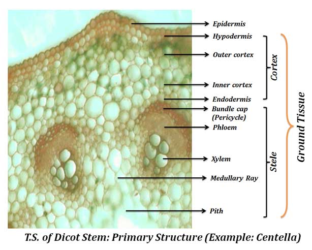
The present post discusses the Primary Structure of Dicot Stem studied under microscope. The anatomy of dicot stem is studied by a T.S. (transverse section) took through the internode of the stem.
Primary Structure of Dicot Stem
Ø The components of cortex and stele are together known as Ground Tissue.
Ø Anatomically the dicot stem has the following regions:
(1). Epidermis
(2). Cortex
a). Hypodermis
b). Outer cortex
c). Inner cortex
d). Endodermis
(3). Stele
a). Pericycle
b). Vascular bundles
c). Medullary rays
d). Pith
(1). Epidermis
Ø Epidermis is the outermost layer, composed of parenchymatous cells.
Ø Usually, epidermis composed of single layer of cells.

Ø Cells are closely packed without any intercellular spaces.
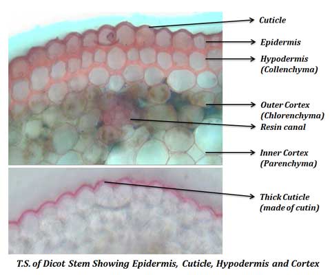
Ø The outer tangential wall of epidermal cells is thicker than other walls.
Ø This wall area is deposited with fatty substances called cutin.
Ø The cutin over the cell wall occurs as separate layer called cuticle.
Ø The epidermis of young stem also contains few stomata.
Ø Multicellular hairs (called trichome) are usually present in the epidermis.
Ø In herbaceous plants, where secondary growth is absent, the epidermis remains throughout the life cycle.
Ø However, in woody plants, the epidermis is replaced after the secondary growth due to back formation.
Ø Functions of epidermis:
o Protection
o Cuticle prevent water loss
o Stomata in stem facilitate gaseous exchange.
o Trichomes and hairs provide protection from fungal spores and insect pests.
(2). Cortex
Ø Cortex is the tissue occupied just inner to the epidermis.
Ø In some plants, the cortex is simple and undifferentiated.
Ø In majority of plants, the cortex is differentiated into many zones.
Ø Usually the cortex in dicot stem composed of FOUR zones.
a. Hypodermis
b. Outer cortex
c. Inner cortex
d. Endodermis
(a). Hypodermis
Ø Hypodermis is the layer of tissue just below the epidermis.
Ø Cells of hypodermis are collenchymatous and with thick primary wall.

Ø Cells are compactly packed without any intercellular space.
Ø In very young stem, the collenchyma is poorly developed.
Ø In stem with ridges and furrows, the collenchyma mainly occurs below the ridges.
Ø Usually, chloroplasts absent in the hypodermis.
Ø Rarely collenchymatous cells of hypodermis do contain chloroplasts.
Ø In xerophytic plants, the hypodermis is sclerenchymatous.
Functions of hypodermis:
o Provide mechanical support.
o In plants with secondary thickening, hypodermal cells give rise to cork cambium which produces the bark.
(b). Outer cortex
Ø Outer cortex consists of the tissue occupied just inner to the hypodermis.
Ø Cells of this region are chlorenchymatous (parenchyma with chloroplasts).
Ø The green colour of young stem is due to his region.
Ø The cells are loosely packed with plenty of intercellular spaces.
Ø In xerophytes, the outer cortical cells forms palisade like tissue for photosynthesis, since these plants usually lack leaves.
Function of outer cortex: photosynthesis
(c). Inner cortex
Ø This is the tissue inner to outer cortex.
Ø Composed of loosely packed parenchymatous cells.
Function inner cortex: storage of carbohydrates.
Special features of cortex in some plants:
Ø In hydrophytes, the cortex is with plenty of air cavities (aerenchymatous).
Ø The Aerenchyma helps in gaseous exchange and provides buoyancy of to plants.
Ø Sclerenchymatous patches occur in the cortex of Eucalyptus, Eugenia, Ficus.
Ø Secretory cavities occur in the cortex of Eucalyptus.
Ø Resin canals occur in the cortex of Anacardium.
Ø Laticifer cells occur in the cortex of latex producing plants.
| You may also like NOTES in... | ||
|---|---|---|
| BOTANY | BIOCHEMISTRY | MOL. BIOLOGY |
| ZOOLOGY | MICROBIOLOGY | BIOSTATISTICS |
| ECOLOGY | IMMUNOLOGY | BIOTECHNOLOGY |
| GENETICS | EMBRYOLOGY | PHYSIOLOGY |
| EVOLUTION | BIOPHYSICS | BIOINFORMATICS |
Functions of cortex
Ø Hypodermal layer provides mechanical support.
Ø During secondary growth, the hypodermal cells give rise to the cork cambium (phellogen) for the bark formation.
Ø Chlorenchymatous cells in the outer cortex can do photosynthesis.
Ø Parenchymatous cells of inner cortex can store carbohydrates.
Ø Cortical cells also store ergastic substances.
Ø Resin canals, latex canals etc. occurs in the cortex.
 (d). Endodermis
 (d). Endodermis
Ø Endodermis is the innermost layer of cortex.
Ø The endodermis is very distinct in lower plants such as Pteridophytes.
Ø NOT distinct in the stem of Gymnosperms and Angiosperms.
Ø Cells of the endodermis accumulate plenty of starch as grains. Thus, the endodermis is also called starch sheath or starch band or starch layer.
Ø If distinct, the endodermis is uniseriate (single layer) with barrel shaped cells.
Ø Cells paranchymatous and they compactly arranged.
Ø Endodermal cells have characteristic thickness in radial and inner tangential walls.
Ø This thickening is called casparian thickening (casparian band, casparian layer).
Ø The casparian band is composed of suberin and lignin, both of them are impervious to water.
Ø Due to the presence of casparian thickening, they block the passage of water and solutes through the protoplasts of endodermal cells.
Functions of endodermis
Ø The exact function of endodermis is not known.
Ø They do not allow the passage of water from cortex to stele, thus may have specific role in the conduction of water.
Ø They can store food material as starch grains.
 (3). Stele
 (3). Stele
Ø Stele is the central vascular cylinder of the stem.
Ø The stele of stem composed of four components.
a) Pericycle
b) Vascular bundle
c) Medullary rays
d) Pith
(a). Pericycle
Ø Pericycle is the outermost layer of the stele.
Ø It is located next (just inner) to the endodermis.
Ø The nature of pericycle in stem shows wide variation.
Ø Pericycle is absent in some plants.
Ø If present, it usually multilayered composed of 3 or more layers of cells.
Ø The pericycle in the stem of different plants may be:
o Completely parenchymatous
o Completely sclerenchymatous
o Mixture of parenchyma and sclerenchyma (alternating bands)
Ø Sclerenchymatous pericycle forms the bundle sheath of the vascular bundle in most of the dicot plants.

(b). Vascular bundle
Ø Vascular bundles (VB) are also called as fascicles.
Ø They are located inner to the pericycle.
Ø VB are developed from the pro-cambium.
Ø The number of vascular bundles is limited in dicot stem.
Ø Usually, 6 to 8 vascular bundles are present and they are arranged as broken ring in the ground tissue.
Ø Vascular bundles of a typical dicot stem are:
o Conjoint: (= xylem and phloem together as bundle)
o Open: (= vascular bundles with cambium)
o Collateral or Bicollateral
Ø Collateral: the usual type of vascular bundle composed of once patch of xylem and one patch of phloem and a strip of cambium between them.
Ø Biocollateral: a special type of vascular bundle composed of a median patch of xylem laying in-between two phloem patches.
Ø Bicollateral VB is characteristic of Cucurbitaceae family (Example: Cephalandra, Cucurbita).
Learn more: Vascular bundles: Structure and Classification
Ø The vascular bundles composed of (I) Xylem placed inner to cambium; and (II) Phloem placed outer to cambium.
 (I). Xylem
Ø Xylem is the water and minerals conducting tissue of vascular bundles.
Ø It is a complex tissue, composed of tracheids, vessels, fibres and parenchyma.
Ø Xylem in the VB is differentiated into:
o Protoxylem
o Metaxylem
Protoxylem
Ø Protoxylem is the first formed part of xylem in the VB.
Ø It is arranged towards the centre of the stem.
Ø Protoxylem composed of very less amount of tracheary elements and large amount of parenchyma.
Ø Tracheary elements are with very narrow lumen.
Ø They show annular or spiral thickening in their secondary wall (primitive type).
Metaxylem
Ø Metaxylem is the xylem part formed after the protoxylem.
Ø It is arranged towards the exterior of the stem.
Ø They composed of more tracheary elements then protoxylem.
Ø The cells of the tracheary elements are with large lumen than that of protoxylem.
Ø They show reticulate or pitted thickening (advanced type).
Ø Functions of xylem:
o Conduction of water
o Conduction of minerals
o Provide mechanical support
o Xylem parenchyma store food materials
(II). Phloem
Ø Phloem is the food conducting tissue of vascular bundles.
Ø Similar to xylem, phloem is also a complex tissue composed of sieve tubes, companion cells, phloem parenchyma and phloem fibres.
Ø The primary phloem is differentiated into:
o Protophloem: first formed phloem, arranged towards periphery.
o Metaphloem: differentiated after protophloem, located near to cambium.
(III). Cambium
Learn more: Characteristics of Meristematic cells
Learn more: Difference between meristem and permanent tissue
Learn more: Classification of Meristems
Ø Cambium is a layer of meristematic tissue present between xylem and phloem.
 Ø Cambium present in the VB is called as fascicular cambium or vascular cambium.
Ø It is the remnant of original pro-cambium.
Ø The cambial cells are parenchymatous and thin primary cell wall.
Ø Cells with dense cytoplasm and prominent nucleus.
Ø Secondary growth in dicots occurs due to the activity of cambium.
Ø Vascular bundle with cambium is called ‘open vascular bundle’.
(c). Medullary Ray
Ø It is also called pith ray.
Ø Medullary ray is a layer of tissue occurs between vascular bundles.
Ø It is composed of loosely packed parenchymatous cells with plenty of intercellular spaces.
Ø The cells of the medullary ray are radially elongated.
Ø During secondary growth, cells of the medullary rays give rise to inter-fascicular cambium.
Ø The fascicular and inter-fascicular cambium fuse together to form a complete ring of cambium and this produce secondary xylem and secondary phloem.
Functions of medullary ray
Ø Allow radial transport of water.
Ø Provide inter-fascicular cambium during secondary growth.
(d). Pith
Ø It is also called as medulla.
Ø Pith is the exact central portion of the stem.
Ø It is located towards the inner side of vascular bundles.
 Ø Usually, the pith composed of parenchymatous cells.
Ø Parenchyma may be loosely arranged with many intercellular spaces.
Ø Sometimes the parenchymatous cells undergo secondary wall thickening.
Ø Cells of outer region of the pith are smaller whereas, those in the inner region larger.
Ø In some plants, the pith is replaced by a large air filled cavity called Pith Cavity.
Function of pith: storage of food materials
Identification reasons of Dicot Stem Primary Structure (Practical exam)
Stem
Ø Vascular bundle conjoint, open, collateral or bicollateral.
Ø Xylem endarch (protoxylem arranged towards the centre).
Ø Endodermis not distinct.
Dicot stem
Ø Collenchymatous hypodermis.
Ø Ground tissue differentiated to hypodermis, cortex and stele.
Ø Limited number of vascular bundles, usually 6 to 8
Ø Vascular bundles are arranged as a broken ring
Ø Vascular bundles, conjoint, open, collateral or bicollateral.
Ø Pith large and well developed.
Reference:
Ø Prakash J.J., 2000, Test Book of Plant Anatomy, Ed. 2, Emkay Publications, New Delhi
Ø Esau K, 1965, Plant Anatomy, Ed. 2, Wiley Eastern Private Limited, New Delhi
Key Questions
Ø The primary structure of a typical dicot stem
Ø Tissue differentiation in dicot stem
Ø Structure of vascular bundle in dicot stem
Ø How dicot stem is different from the monocot stem?
Ø How stem is different from root?
Ø What are the functions of medulla and pith?
Ø Differentiate collateral and bicollateral vascular bundles
Ø What is the importance of casparian thickening?
| You may also like... | ||
|---|---|---|
| NOTES | QUESTION BANK | COMPETITIVE EXAMS. |
| PPTs | UNIVERSITY EXAMS | DIFFERENCE BETWEEN.. |
| MCQs | PLUS ONE BIOLOGY | NEWS & JOBS |
| MOCK TESTS | PLUS TWO BIOLOGY | PRACTICAL |
Proud of you. Excellent notes .
Plz how to download botany notes in ppt or in pdf?plz guide me
Thank you, keep visiting easybiologyclass