
Phase Contrast vs DIC
Phase Contrast and Differential Contrast Microscopes
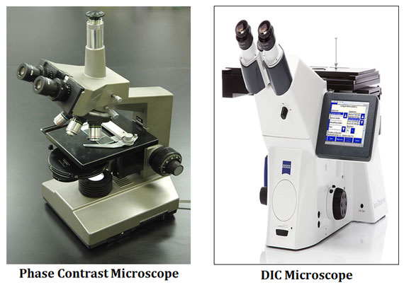
Phase contrast microscopy and Differential Interference Contrast (DIC) microscopy are two advanced optical light microscopy techniques to produce high contrast images of unstained and living cells. Both the microscopes utilize various contrast enhancing techniques to produce high contrast images.
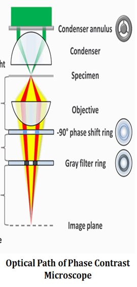
Phase contrast microscopy is an optical-microscopy technique developed by Frits Zernike in 1934 to produce high contrast images of unstained live specimens. The phase contrast microscopy works by converting the phase shifts of light passing through a transparent specimen to detectable brightness changes in the image.
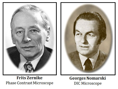
Differential Interference Contrast (DIC) microscopy, also called as Nomarski Interference Contrast (NIC) Microscopy, was first invented by Georges Nomarski in 1952. DIC microscopy uses more sophisticated contrast enhancing techniques than phase contrast system. It works by separating a polarized light source into two orthogonally polarized mutually coherent parts which are spatially displaced at the sample plane, and recombined before the final image formation. DIC produce more pronounced contrast difference than phase contrast image.
| You may also like NOTES in... | ||
|---|---|---|
| BOTANY | BIOCHEMISTRY | MOL. BIOLOGY |
| ZOOLOGY | MICROBIOLOGY | BIOSTATISTICS |
| ECOLOGY | IMMUNOLOGY | BIOTECHNOLOGY |
| GENETICS | EMBRYOLOGY | PHYSIOLOGY |
| EVOLUTION | BIOPHYSICS | BIOINFORMATICS |
The present post discusses the similarities and differences between phase contrast microscopy and Differential Interference Contrast Microscopy with a Comparison Table.
Similarities between Phase Contrast Microscopy and DIC Microscopy
Ø Both Phase Contrast and DIC microscopes produce high contrast images.
Ø Both are light microscopes, uses the visible spectrum of light.
Ø Both uses two light beams to generate the final image.
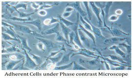
Ø Both are used for visualizing the live cells and living events.
Ø Staining is not required for both types of microscopes.
Ø Both the microscopes follow the Abbe’s law (limit of resolution).
Difference between Phase Contrast Microscopy and DIC Microscopy
Sl. No. Phase Contrast Microscope (PCM) Differential Interference Contrast Microscope (DIC)

1 Phase contrast microscopy was developed by Zernike in 1934 DIC was developed by Nomarski in 1952
2 Phase contrast microscope works by the detection of sharp changes in the refractive index DIC microscope works by the detection of continuous change of refractive index
3 Phase contrast microscopy utilizes two light beams without beam splitters DIC utilizes two light beams with beam splitters

4 Produce image by the incomplete separation of direct and diffracted light rays Produce image by the complete separation of direct and diffracted light rays
5 Does not use a polarizer Uses a polarizer
6 Optical system of phase contrast microscopy do not requires beam recombiness Optical system of DIC requires beam recombiners
7 Uses annular diaphragm and phase plate to create contrast Does not use annular diaphragm and phase plate
8 Nomarski’s prism is not used in the optical path Nomarski’s prisms are used in the optical path
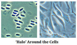
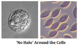
9 Produce a ‘halo’ around edges of the image No ‘halo’ is produced around the edges on the image
10 Images from the phase contrast microscopy only provide qualitative data Images from the DIC can produce both qualitative and quantitative data
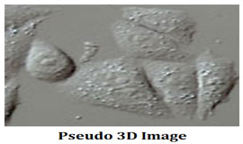
11 No three dimensional effects on the images Images are with a pseudo three dimensional effect

12 Full numerical aperture of the condenser and objective lenses cannot be utilized because of the masking effect of phase plate used in the light path DIC microscopes can utilize the full numerical aperture of objective and condenser lenses
13 Images in phase contrast microscope are not sensitive to the orientation of specimen Images in the DIC are highly sensitive to the orientation of the specimen
14 Axial resolution low Axial resolution high
15 Optical sectioning is not possible Optical sectioning is possible due to the high axial resolution
16 Cost of the microscope is moderate Cost is very high
You might also like…
@. Phase Contrast Microscope- Working Principle
@. Difference between Light Microscope and Electron Microscope