
Gram Positive vs Gram Negative Bacteria
What is Grams staining?
Christian Gram, a Danish Physician in 1884 developed a staining technique to distinguish two types of bacteria. The two categories of bacteria based on gram staining are Gram positive bacteria and Gram negative bacteria. Bacteria are first stained with crystal violet or gentian violet. All bacterial cells will stain blue or purple colour with crystal violet solution. Then the bacterial cells are treated with iodine solution (Lugol’s iodine) solution and washed with alcohol (de-staining solution). Those bacteria which retain the blue or purple colour of crystal violet are called Gram positive bacteria and those bacteria which loose the colour of crystal violet after washing with de-staining solution is called Gram Negative bacteria.
Gram negative bacteria are later stained with safranin or fuchsin for observation under microscope. Gram negative bacteria after safranin or fuchsin staining will appear red or pink colour. Gram staining differentiates bacteria by the chemical and physical properties of their cell walls by detecting the properties of peptidoglycan. Gram staining method is useful in differentiating majority of bacterial species into two broad categories. Even though all bacterial species cannot be differentiated based on gram staining technique, this method has immense application in clinical diagnostics and biological researches.
Similarities between Gram Positive and Gram Negative Bacteria
Ø Both are bacterial cells
Ø Both groups are prokaryotic
Ø Both lack membrane bounded organelles
Ø Both groups have covalently closed circular DNA as the genetic material
Ø Both groups contain extra-chromosomal genetic materials (plasmids)

Gram Positive (blue) and Gram Negative (Pink) Bacteria (source wikipedia)
Ø Both groups possess capsule
Ø In both groups, cell wall is made up of peptidoglycan
Learn more: Peptidoglycan vs Pseudo-peptidoglycan
Ø In both groups, cytoplasm is surrounded by lipid bilayer with many membrane spanning proteins
Ø Both gram-positive and gram-negative bacteria commonly have a surface layer called an S-layer
Ø Both groups of bacteria undergo genetic recombination through transformation, transduction and conjugation
Ø Both groups undergo binary fission as a mode of asexual reproduction
Ø Both groups contain many flagellated and non-flagellated species
Learn more: Cell Surface Appendates Flagella vs Fimbriae vs Pili
Ø Both gram positive and gram negative bacteria are inhibited by antibiotics (their sensitivity varies)
Ø Both groups includes flagellated (motile) and non-flagellated (non-motile) forms
Learn more…
Gram Positive Cell Wall vs Gram Negative Cell Wall
Difference between Prokaryotes and Eukaryotes
Difference between Archaebacteria, Bacteria and Eukaryote
Difference between Prokaryotic and Eukaryotic Transcription
Difference between Prokaryotic and Eukaryotic Translation
Difference between Gram Positive Bacteria and Gram Negative Bacteria
Sl. No. Gram Positive Bacteria Gram Negative Bacteria
1 Appears as dark violet or purple coloured under microscope after Gram staining Appears as pink or red coloured after Gram staining
2 Retain the colour of crystal violet after washing with alcohol (de-staining solution) Colour of crystal violet will not be retained after washing with de-staining solution


3 Examples: Bacillus subtilis, Streptococcus, Lactobacillus, Leuconostoc, Clostridium Examples: Escherichia coli, Rhizobium, Vibrio, Acetobacter
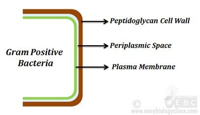
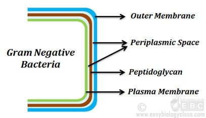
4 Cell wall single layered, straight and even Cell wall two layered and uneven (wavy)
5 Cell wall is very rigid and less elastic Cell wall is less rigid and more elastic
6 The rigidity of cell wall is due to the high proportion of peptidoglycans (80%) The elasticity of cell wall is due to the less amount of peptidoglycan (2 – 12 %)
7 Thickness of cell wall varies from 15- 20 nm, sometimes up to 80 nm Thickness of cell wall varies from 7.5 to 12 nm
8 Muramic acid content of cell wall is more, 16 – 20% of dry weight Muramic content of cellwall is less, 2 – 5% of dry weight
9 Cell wall is resistant to alkalis and insoluble in 1% KOH solution Cell wall is sensitive to alkalis and soluble in 1% KOH solution
10 Cell is highly resistant to physical disruptions Cell is highly susceptible to physical disruptions
11 S-layer is attached to the peptidoglycan layer S-layer is attached to the outer membrane
12 Teichoic acid present in the cell wall Teichoic acid absent in the cell wall
13 Periplasmic space absent or if present, very narrow Periplasmic space present
14 Outer membrane absent Outer membrane present
15 Lipid content very low (1-4%) Lipid content very high (11-22%)
16 Porins (proteinaceous membrane channels) are absent Porins are present
17 Lipopolysaccharides usually absent Lipopolysaccharides present
18 Mesosomes are more prominent Mesosomes are less prominent
19 Produce endospores spores during unfavourable conditions Usually do not produce endospores
20 Flagella with 2 rings in the basal body Flagella with four rings in the basal body
21 Usually produce exotoxins Usually produce endotoxins
22 Gram positive bacteria are more susceptible to antibiotics Gram negative bacteria are more resistant to antibiotics
23 Can be killed by vancomycin antibiotic Cannot be killed by vancomycin antibiotic
24 Shows high resistance to sodium azide solution Shows low resistance to sodium azide solution
25 Cells shows high susceptibility towards penicillins and sulfonamide antibiotics Cells shows low susceptibility towards penicillins and sulfonamide antibiotics
26 Cells show low susceptibility towards streptomycin, chloramphenicol and tetracyclines Cells shows high susceptibility towards streptomycin, chloramphenicol and tetracyclines
27 Cell wall is highly susceptible to degradation by lysozyme enzyme Cell wall is less susceptible to degradation by lysozyme enzyme
28 Shows high tolerance towards dryness Shows low tolerance towards dryness
29 Magnetosomes usually absent Magnetosomes sometimes present
30 Very few are pathogenic to human and other animals Most of them are pathogenic to human and other animals