
The Folding Mystery of Chromosomes
The chromosomes of eukaryotic organisms are a complex structural organization of DNA and proteins. The exact structural organization of proteins and DNA to form the chromatin material (or chromosomes during cell division) is a curious question in the scientific community. This curiosity becomes a wonder when we realize the total length of DNA in a single cell and size of the nucleus in which this DNA is residing.
For example, in a diploid human cell, there will be 46 chromosomes. The DNA in all these 46 chromosomes when joined together, it will have a distance of about 2.2 meters. Thus, the average length of DNA in a single chromosome will be 4.8 cm or 48,000 µm (2.2 X 100/46). On an average, the human chromosome at its metaphase stage is about 6 µm long. This means the 48,000 µm long DNA strand is heavily folded to from the 6 µm long chromosome with a packing ratio of about 8000 : 1. The exact folding pattern of DNA is a highly debated concept.
| You may also like NOTES in... | ||
|---|---|---|
| BOTANY | BIOCHEMISTRY | MOL. BIOLOGY |
| ZOOLOGY | MICROBIOLOGY | BIOSTATISTICS |
| ECOLOGY | IMMUNOLOGY | BIOTECHNOLOGY |
| GENETICS | EMBRYOLOGY | PHYSIOLOGY |
| EVOLUTION | BIOPHYSICS | BIOINFORMATICS |
For explaining the structural organization of DNA and proteins in the chromosome, various theories have been put forward by different scientists. DuPraw Folded Fibre Model and Nucleosome Model are the two such models trying to explain the ultra-structural organization of DNA and proteins in the chromosome. The present post describes the significance of Folded Fibre Model of Chromosomes and its merits and demerits.
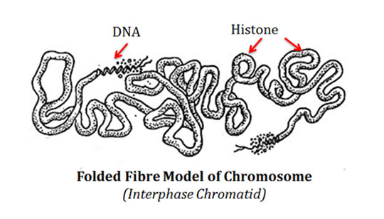
Folded Fibre Model of Chromosome
Ø The Folded Fibre Model of chromosome was proposed by DuPraw in 1965.
Ø He published this model based on his studies on human chromosomes using electron microscope.
Ø The important features of Folded Fibre Model of Chromosome are:
@. Each chromosome contains a single but long and coiled chromatin fibre.
@. This single chromatin fibre is more or less uniform in thickness.
@. He suggested the thickness of this fibre around 200 to 250 Å.
@. This single chromatin fibre contains a single DNA double helix with associated proteins.
@. The 20Å thick DNA double helix is packed spirally to form a fibre.
@. This fibre is then coiled to form a 10 to 100 Å fibres called type A fibre.
@. The packing ratio of DNA in type A fibril is about 6 : 1.
@. Type A fibre further coiled to form 200 to 250 Å thick type B fibre with a folding ratio of 10 : 1.
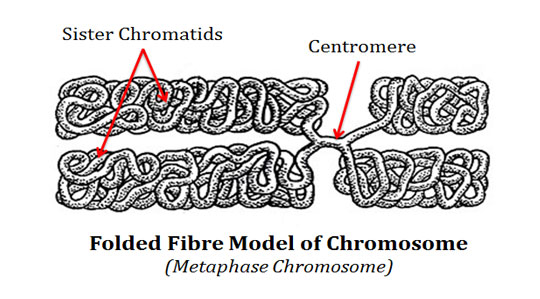
@. The 200-250 Å type B fibre is further folded to form the chromatid.
@. This fiber is supposed to contain DNA histone helix.
@. This DNA histone helix is in a super coiled condition.
@. The histone protein bound on outer side of DNA and forms a shell around the DNA.
@. DuPraw called this histone coat of chromosome as the ‘histone shell’.
@. In the S phase of cell cycle, the chromatid undergo a round of replication to form two sister chromatids.
@. The sister chromatids are held together by an unreplicated region of the chromatin fibre.
| You may also like... | ||
|---|---|---|
| NOTES | QUESTION BANK | COMPETITIVE EXAMS. |
| PPTs | UNIVERSITY EXAMS | DIFFERENCE BETWEEN.. |
| MCQs | PLUS ONE BIOLOGY | NEWS & JOBS |
| MOCK TESTS | PLUS TWO BIOLOGY | PRACTICAL |
Demerits of Folded Fiber Model of Chromosome
Ø DuPraw’s Folded Fibre Model of chromosome is not accepted in the scientific community.
Ø The DNA-histone association as described in this model is now considered unlikely by the discovery that DNA itself is looped around histone beads to form nucleosomes.
Ø We now know that histones are not forming a shell around DNA.
Ø The DNA is not buried inside a protein shell.
Ø DNA is freely exposed along the entire surface for genetic expression.
Ø DuPraw model was replaced by the most popular and universally accepted Nucleosome model of chromosome.
<<Back to Cell & Molecular Biology Lecture Notes