
What is Apoptosis? Why apoptosis is known as the ‘Programmed Cell Death’?
The total number of cells in an organ or organism is fundamentally fixed to a specific range in all multicellular organisms. In every multi-cellular organism, the cell number is effectively controlled by two strategies- (a) by regulating cell Division and (b) by regulating cell Death. If cells are no longer needed, they commit suicide (self-destruction) by activating an intracellular death signaling programme. Thus, this death process is known as ‘Programmed Cell Death’. This programmed cell death pathway is called Apoptosis.
The term apoptosis in Greek literally mean ‘falling off’. Just like the old leaves ‘falloff’ from the trees without affecting the life of the plant, the apoptotic cell death will not interfere with the functioning of the organ and organism. The most striking feature of apoptosis is that if a cell undergoes the programmed cell death, the neighboring cells are not at all damaged. Apoptotic death of a cell and its subsequent phagocytosis by a neighboring cell or by a macrophage allow the organic components of the death cell to be effectively recycled.
Learn more: Difference between Apoptosis and Necrosis
The apoptosis is better known as the ‘Programmed Cell Death’. It is a natural well-orchestrated, well sequenced and timely executed chain of events leads to the death of a cell.
| You may also like NOTES in... | ||
|---|---|---|
| BOTANY | BIOCHEMISTRY | MOL. BIOLOGY |
| ZOOLOGY | MICROBIOLOGY | BIOSTATISTICS |
| ECOLOGY | IMMUNOLOGY | BIOTECHNOLOGY |
| GENETICS | EMBRYOLOGY | PHYSIOLOGY |
| EVOLUTION | BIOPHYSICS | BIOINFORMATICS |
What are the characteristics of Apoptotic Cell Death?
An apoptotic cell death is characterized by:
Ø Shrinkage of the cell
Ø Shrinkage of the nucleus
Ø Loss of adhesion to the neighboring cells
Ø Formation of membrane blebs (externalization of inner leaflet of membrane)
Ø Decay of mRNA
Ø Condensation and fragmentation of the chromatin (DNA)
Ø Formation of small fragmented chromatin in membrane bounded structures called apoptotic bodies
Ø Rapid engulfment of the apoptotic cell debris by the process of phagocytosis
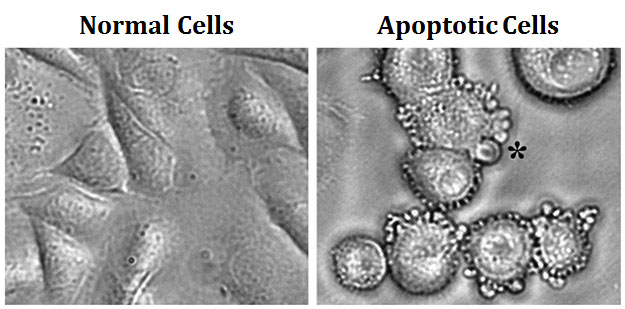
image source: cc wikipedia
What are ‘apoptotic bodies’?
During apoptotic cell death, the nucleus gets fragmented into many discrete chromatin bodies due to degradation of nuclear DNA. Each such nuclear fragment is surrounded by blebbed plasma membrane and these units were bud-off from the apoptotic cell. Thus, by the completion of apoptosis, the cell content is converted into many small vesicles called ‘apoptotic bodies’. Apoptotic bodies are immediately phagocytosed by the macrophages or surrounding healthy cell.
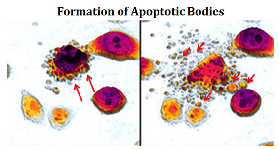
(image source: cc wikipedia)
Does apoptosis be Natural or Pathogenic?
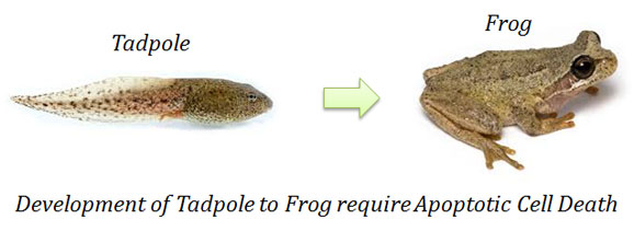
The apoptosis is a natural process. About 1010 to 1011 cells in the human body dies every day by the process of apoptosis. Apoptosis is essential for the proper embryonic development in higher organisms. For example, the separation of fingers and toes in a developing human embryo occurs because cells between the digits undergo apoptosis during the embryonic development. Apoptosis also helps to prevent the perpetuation of lethal genetic damages in the body. The apoptotic cell death can occur in a cell when its genetic material is severely damaged and it cannot be rectified by the inbuilt DNA repair mechanism. Sometimes, the apoptosis can be pathogenic such as the death of healthy neurons which leads to the Alzheimer’s disease.

Webbed toes formation due to the lack of apoptotic cell death during embryonic development (cc wikipedia)
How was apoptosis discovered?
The term apoptosis was coined by John Kerr, Andrew Wyllie and A.R. Currie in 1972. The molecular basis of apoptosis was elucidated for the first time by the studies in a nematode Caenorhabditis elegans. The worm C. elegans constantly maintain their cell number in its embryonic and adult stages. During the embryonic development, the worm produces exactly 1090 cells. Among these 1090 cells, 131 cells are precisely destined to die by apoptosis during the development. Further studies in the worm identified a specific gene involved in controlling the apoptosis process and it is named as CED-3. A worm with inactive CED-3 gene by mutation fails to induce the apoptotic cell death in the embryonic development stage. This shows that CED-3 plays a crucial role in executing the process of programmed cell death. Later, scientists identified genes homologous to the CED-3 of C. elegans in other organisms including humans and subsequently named as Caspases.

Caenorhabditis elegans (image source: cc Wikipedia)
What are caspases? What is the importance of caspases in apoptosis?
Caspases are a family of proteins present in human and other animals which are homologous to the CED-3 gene product of C. elegans. Caspases are cysteine proteases involved in the execution of apoptotic cell death. Cysteine proteases are a category of protease enzymes with a cysteine residue at its active site. The caspases are produced as inactive zymogens called pro-caspases. Pro-caspases are activated to caspases during the early stages of apoptosis. Activation of pro-caspase to caspases is achieved by the catalytic removal of a part of the peptide chain. Activated caspases are responsible for most of the molecular events in the apoptosis signaling pathway.
How caspases execute apoptosis? What are the targets of caspases during apoptosis?
Caspases execute the apoptosis by selectively targeting and cleaving a large array of key molecules in the cells. Most important target molecules of caspases during apoptosis are given below:
(1). Protein Kinases: Protein kinases such as Focal Adhesion Kinase (FAK), Protein Kinase B (PKB), Protein Kinase C (PKC) and Raf1. Inactivation of the FAK cause detachment of the apoptotic cells from its neighboring cells due to the inhibition of cell adhesion.
(2). Lamin: Lamin form the inner lining of the nuclear membrane and thus the cleavage of lamins lead to the disintegration of nuclear lamina (nuclear membrane) and breakage of the nucleus.
(3). Cytoskeleton proteins: The cleavage of cytoskeleton proteins such as actin, tubulin and intermediate filaments lead to the shrinkage of the cells.
(4). CAD (Caspase Activated DNases): CAD is an endonuclease. In a normal cell, the CAD endonuclease exists in an inactive stage. The cleavage of CAD by caspase activates the CAD enzyme. Activated CAD then translocated into the nucleus and it cleaves and degrades the DNA.
What are apoptotic signals?
Any stimuli that can induce and initiate the programmed cell death pathway are called apoptotic signals. The source of apoptotic signals can be of two different types such as those from the external sources and those signals originated in the cell itself. Based on the source of signals, there are essentially two types of apoptotic signaling pathways. They are:
(1). Intrinsic Apoptotic Pathway: Here the apoptotic stimuli are originated internally in the target cell itself. The most important internal signal that induces intrinsic signaling is severe DNA damage that cannot be rectified by the DNA repair mechanism.
(2). Extrinsic Apoptotic Pathway: Here the stimuli are from the external source (not from the cell itself). The most important external apoptotic signals are cytokines such as Tumour Necrosis Factor (TNF). (The exact mechanism of intrinsic and extrinsic pathways of apoptosis will be discussed later).
Learn more: Intrinsic Pathway of Apoptosis
Learn more: Extrinsic Pathway of Apoptosis
Even though the signaling cascades of extrinsic and intrinsic pathways are separate, there is always cross-talk between these two pathways. The extrinsic pathway can induce the activation of the intrinsic pathway of apoptosis.
| You may also like... | ||
|---|---|---|
| NOTES | QUESTION BANK | COMPETITIVE EXAMS. |
| PPTs | UNIVERSITY EXAMS | DIFFERENCE BETWEEN.. |
| MCQs | PLUS ONE BIOLOGY | NEWS & JOBS |
| MOCK TESTS | PLUS TWO BIOLOGY | PRACTICAL |
What are the significances of apoptosis?
Apoptosis is a beneficial event. Moreover, failure to regulate apoptosis can result in the damage of organs or organisms. The main significances of apoptosis are given below:
Ø Apoptosis help to maintain the homeostasis in multicellular organisms.
Ø Apoptosis also helps to maintain the proper body size.
Ø Apoptosis maintains the constancy of cell number in an organ or organism.
Ø Apoptotic cell death is a pre-request for the proper embryonic development.
Ø By the process of apoptosis, the body can eliminate unwanted cells such as:
$ A cell with severely damaged DNA
$ A cell with fatal mutation
$ A pathogen (virus) infected cell
$ Unwanted cells formed during embryonic development
$ Cells that to be killed during proper neuronal architecture development
Ø Apoptosis also helps to kill T lymphocytes with receptors for the proteins present on the normal cell. These T cells are produced during the embryonic development. These dangerous T lymphocytes are eliminated by apoptotic cell death.
Ø Apoptotic cell death can be pathogenic in some cases.
Ø Apoptosis is involved in some neurodegenerative diseases such as Alzheimer’s, Parkinson’s disease, and Huntington’s disease by the elimination of essential neurons.
Ø Failure to induce apoptosis is the main reason for most of the cancers.
How the macrophages specifically recognize the apoptotic cells for phagocytosis?
Both the intrinsic and extrinsic pathway of apoptosis converges by activating the same executioner caspases, i.e., caspase-3. As the apoptotic signaling proceeds, the cell loses its contact with the neighboring cell and starts to shrink. The cell ultimately shrinks into one or more condensed membrane-enclosed structures called the apoptotic body. The apoptotic bodies are characterized by the presence of phosphatidyl serine on their outer surface. Phosphatidyl serine is a membrane lipid present only in the inner leaflet of plasma membrane. During apoptotic cell death, the plasma membrane flipping occurs which results in the externalization of phosphatidyl serine residues. These externalized phosphatidyl serine molecules are the ‘eat me signals’ for the macrophages. The macrophages recognize these ‘eat me’ signal and they completely phagocytosis the apoptotic bodies. Thus, the apoptotic cell death is completed without spilling the cellular content into the extracellular environment. This is very significant because the release of cell debris can trigger inflammatory responses which ultimately cause severe tissue damage.
What is the relationship between Apoptosis and Cancer?
Cancer is a pathological process of uncontrolled division of cells leading to tumor development. Some cancerous cells also have the potential to invade healthy tissues by a process called metastasis. The cancer is essentially the uncontrolled division of an abnormal cell with mutations of genetic damage. If the apoptotic signaling is properly working, these unwanted cells can be eliminated from the body by programmed cell death pathway. Thus the main reason for cancer is the failure to induce apoptosis in an unwanted cell and as a result of this, the unwanted cell perpetuates without any control.
Learn more: Cell Cycle Checkpoints and Regulation of Cancer (short notes)
Review Questions…
(1). Define Apoptosis.
(2). Why is apoptosis known as the ‘Programmed Cell Death’?
(3). What are the characteristics of Apoptotic Cell Death?
(4). What are apoptotic bodies?
(5). Does apoptosis be Natural or Pathogenic?
(6). What is meant by membrane blebbing?
(7). How was apoptosis discovered?
(8). What are caspases? What is the importance of caspases in apoptosis?
(9). How caspases execute apoptosis? What are the targets of caspases during apoptosis?
(10). What are apoptotic signals?
(11). What are the significances of apoptosis?
(12). What is the relationship between Apoptosis and Cancer?
(13). What is the difference between apoptosis and necrosis?
(14). What is the importance of apoptosis in embryonic development?
(15). What is the role of C. elegans in the discovery of apoptosis?
(16). Who discovered apoptosis?
(17). What is CED? What is its importance in apoptosis?
(18). What are the main targets of caspases in human during apoptosis?
(19). What is meant by intrinsic pathway of apoptosis?
(20). What is meant by extrinsic pathway of apoptosis?
(21). Give some examples of intrinsic apoptotic signals.
(22). Give some examples of extrinsic apoptotic signals.
(23). Differentiate pro-caspases and caspases.
<<Back to Molecular Biology Notes