
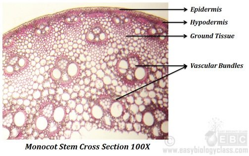
The anatomy or internal structure of a monocot stem can be studied by a Transverse Section (T.S.) taken through the internode of a monocot plant such as grass, bamboo, maize, Asparagus etc. The main difference of monocot stem from dicot stem is that, here in monocots the ground tissue is NOT differentiated into Cortex and Endodermis. The anatomical features of a typical monocot stem are summarized as key points below.
Anatomy of Monocot Stem
@. The T.S. of a monocot stem is usually circular in outline
@. Typically a monocot stem consist of FOUR tissue systems.
(1). Dermal tissue system
(2). Hypodermal tissue system
(3). Ground tissue system
(4). Vascular tissue system
(1). Dermal tissue system
@. Dermal tissue system constitute the epidermis
@. Epidermis forms the outermost layer
@. Usually the epidermis is single layered and made up of parenchymatous cells
@. Epidermal cells are compactly packed without any inter-cellular spaces
@. A thick layer of cuticle is present over the outer wall of epidermis
@. A special feature of monocot epidermis is that, cell wall is highly silicified (they shows silica deposition)
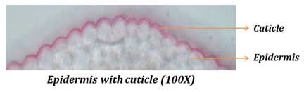
@. Epidermal hairs or trichomes are usually absent in monocots
@. Stomata are present (few in number) on the epidermis
@. Since there is no secondary growth in monocots, the epidermis persists as long as the stem (till the death of the plant)
@. Functions of epidermis
&. Act as the limiting layer and thus forms the outer boundary
&. Provide protection
&. Silica deposition provide mechanical support
&. Cuticle prevent transpiration
&. Stomata present on epidermis allows gaseous exchange
(2). Hypodermal tissue system
@. Hypodermal tissue system consists of hypodermis, it occupies immediately below the epidermis
@. Cells are polygonal and compactly packed without any inter-cellular spaces
@. Hypodermis is multilayered and sclerenchymatous
@. Alternate patches of chlorenchyma may present in some plants (Grass)
@. Below the chlorenchyma, the hypodermis is continuous
@. Peripheral vascular bundles are sometimes embedded in the hypodermis
@. Functions of hypodermis:
&. Provide mechanical support and protection
&. Chlorenchyma can do photosynthesis
(3). Ground tissue system
@. Ground tissue composed of cortex only (Ground tissue NOT differentiated)
@. Cortex is parenchymatous; cells are larger and circular
@. Cortical cells are loosely packed with plenty of inter-cellular spaces
@. In aquatic monocots (Potamogeton) aerenchyma is present in the ground tissue
@. In some plants, central portion of ground tissue is hollow and filled with air (Oryza, Triticum)
@. Functions of ground tissue:
@. Storage of food materials
@. Harbor vascular bundles
| You may also like NOTES in... | ||
|---|---|---|
| BOTANY | BIOCHEMISTRY | MOL. BIOLOGY |
| ZOOLOGY | MICROBIOLOGY | BIOSTATISTICS |
| ECOLOGY | IMMUNOLOGY | BIOTECHNOLOGY |
| GENETICS | EMBRYOLOGY | PHYSIOLOGY |
| EVOLUTION | BIOPHYSICS | BIOINFORMATICS |
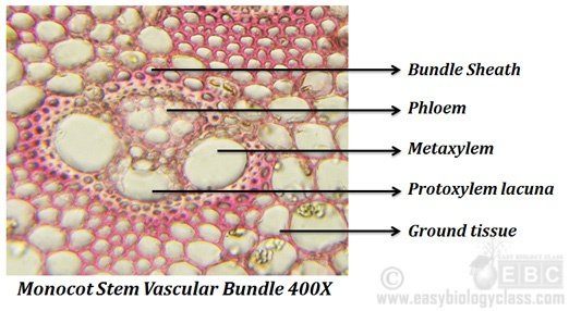
(4). Vascular tissue system
@. Vascular tissue system composed of vascular bundles (VB)
@. Vascular bundles are numerous and they scatteredly arranged in the ground tissue
Learn more: Vascular Bundles- Structure and Classification
@. Vascular bundles are widely separated from each other
@. They shows size differences
@. Vascular bundles in the outer region are smaller, whereas those in the inner region are larger
@. Vascular bundles in most cases are surrounded by a sclerenchymatous bundle sheath
@. Bundle sheath is absent in Asparagus
@. Bundle cap is absent in monocots
@. Vascular bundles are conjoint, collateral and closed
@. Cambium is absent (closed VB) and hence no secondary thickening in monocots
@. Vascular bundles composed of (A). Xylem and (B). Phloem
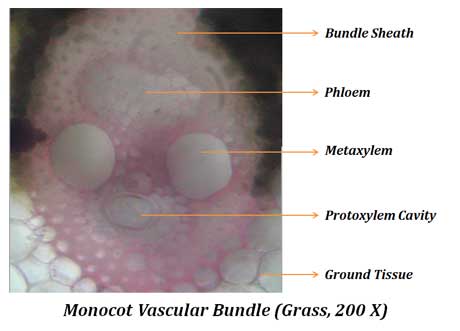
(A). Xylem
@. Xylem is endarch (Protoxylem arranged towards interior and Metaxylem towards exterior)
@. Xylem elements are arranged in the shape of a ‘V’
@. The metaxylem elements forms the arms of ‘V’, whereas the protoxylem elements occupy at the junction of ‘V’
@. Metaxylem composed of only two large vessels with pitted thickening
@. Meta-xylem tracheids present between meta-xylem vessels
@. Protoxylem composed of few vessels with annular or spiral thickening
@. Protoxylem elements fused to form a lysigenous cavity called protoxylem lacuna or protoxylem cavity
@. Protoxylem cavity is absent in Asparagus
@. Function of xylem: conduction of water, provide mechanical support
(B). Phloem
@. Phloem lies outer to xylem (between the two arms of ‘V’)
@. Phloem composed of sieve tubes and companion cells
@. Phloem parenchyma is absent in monocot vascular bundles
&. Function: Conduction of food
Monocot Stem Identification Reasons (Practical)
(1). In T.S., the stem is circular in outline
(2). Epidermis is single layered with thick cuticles, epidermal hairs are absent
(3). Silica deposition in epidermis
(4). Hypodermis is multilayered and sclerenchymatous
(5). Ground tissue is undifferentiated with loosely packed parenchymatous cells
(6). Endodermis and pericycle are absent
(7). Vascular bundles are numerous, scatteredly arranged in the ground tissue
(8). Vascular bundles are conjoint, collateral and closed
(9). Bundle sheath is present, bundle cap is absent
(10). Xylem endarch with two prominent and large metaxylem vessels
(11). Protoxylem forms a protoxylem cavity (lacuna)
(12). Phloem situated outer to the xylem, between the two metaxylem vessels
(13). Central portion hollow or solid
(14). Secondary thickening absent
| You may also like... | ||
|---|---|---|
| NOTES | QUESTION BANK | COMPETITIVE EXAMS. |
| PPTs | UNIVERSITY EXAMS | DIFFERENCE BETWEEN.. |
| MCQs | PLUS ONE BIOLOGY | NEWS & JOBS |
| MOCK TESTS | PLUS TWO BIOLOGY | PRACTICAL |
Please provide the link for monocot leaf also