
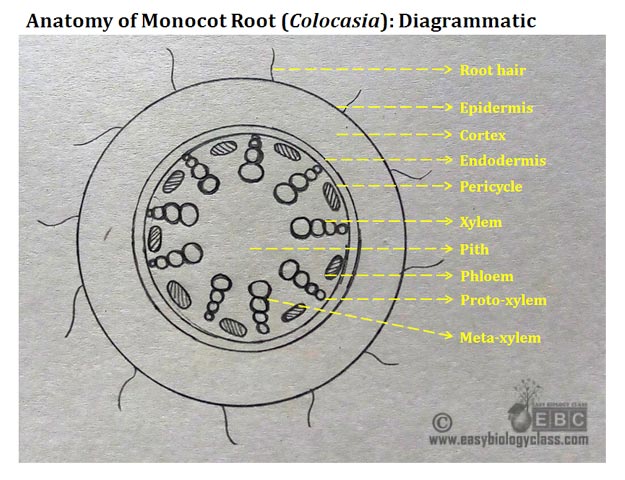
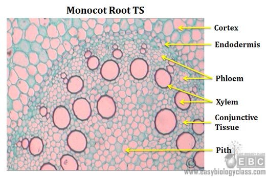
The anatomical features of a monocot root can be studied through a cross section (CS) through the root. The present post discesses the anatomy of monocot root with Monocot Root Diagram.
Ø Anatomically, the monocot root has been differentiated into the following parts:
(1). Epidermis
(2). Cortex
(3). Endodermis
(4). Pericycle
(5). Vascular Tissue
(6). Conjunctive Tissue
(7). Pith
(1). Epidermis
Ø Epidermis in the root is also known as epiblema, piliferous layer and rhizodermis.
Ø It is the outermost layer in the root, composed of closely packed parenchymatous cells.
Ø Usually, the epidermis is single layered with thin walled cells.
Ø Cuticle is absent. Cuticle is present in some aerial roots.
 Ø Many epidermal hairs, called root hairs, are present on the epidermal cells.
Ø Root hairs are unicellular, un-branched epidermal cell extensions with very thin cell wall.
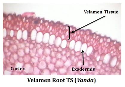
Ø In Orchids, the epidermis is multilayered and transformed into specialized water absorbing Velamen Tissue.
Ø Cells of the velamen tissue are dead with specialized ‘band’ like thickenings.
Ø Velamen tissue absorbs water moisture from the atmosphere.
Ø They also provide mechanical support and prevent the water loss from cortex.
Functions of Epidermis
o Epidermis acts as the outermost boundary.
o Root hairs help to absorb water and nutrients from the soil.
o Velamen tissue in Orchids helps to absorb water from atmosphere.
(2). Cortex
Ø Cortex is the ground tissue occupies between the epidermis and the vascular tissue.
Ø Cortex is well developed in monocot roots.
Ø Composed of loosely packed parenchymatous cells with plenty of intercellular spaces.
Ø In Zea mays the outer cortical cells just below the epidermis are thick walled (called hypodermis).
Ø In some plants, the cortex is very large and differentiated into three zones.
Ø Outer cortex: composed of small thin walled cells.
Ø Middle cortex: composed of large cells which serve as storage space.
Ø Inner cortex: composed of few large and thin walled parenchymatous cells.
Ø In some aquatic plants (Oryza sativa, Potamogeton) the cortex is traversed by large air spaces or lacunae. Such cortex is called lacunate cortex.
Functions of cortex
o Cortical cells can do photosynthesis in aerial roots.
o Aerenchyma in cortex allows gaseous exchange.
o Aerenchyma in cortex provides buoyancy in aquatic plants.
o Cortical cells can store starch and ergastic substances.
o Cortex also harbours resin canals and ducts.
 (3). Endodermis
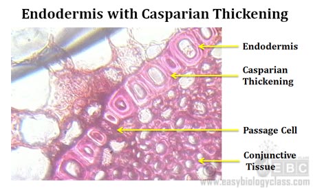
Ø Endodermis is the innermost layer of cortex.
Ø Endodermis is prominent and conspicuous in roots.
Ø Cells of endodermis are large and barrel shaped.
Ø They possess special thickening in their radial and inner tangential walls.
Ø Such thickening is called Casparian thickening or Casparian band.
Ø The endodermal cells opposite to the proto-xylem remain thin walled (they lack the casparian thickening).
Ø These thin walled endodermal cells are called Passage Cells.
Ø Passage cells allow the transport of water from the cortex to the xylem elements.
Ø The endodermal cells store plenty of starch grains and hence it is called starch sheath.
Functions of endodermis
o The exact function of endodermis is not known.
o Casparian thickening in the endodermis does not allow the free passage of water from cortex to the stele.
o Endodermal cells can store food material as starch grains.
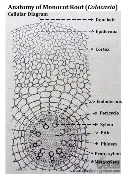
Monocot Root Diagram
 (4). Pericycle
Ø The layer(s) of cells occupying between the endodermis and vascular tissue is called pericycle.
Ø Usually, the pericycle composed of a single layer of thin walled parenchymatous cells.
Ø In Smilax, the pericycle is multiseriate and thick walled
Functions of pericycle
o Lateral roots originate from pericycle
(5). Vascular Tissue
Ø Vascular tissue consists of alternating strands of xylem and phloem (radial arrangement).
Ø Number of xylem and phloem strands varies from 6 to many (hexarch to polyarch).
Ø Xylem is exarch (proto-xylem oriented towards the exterior, meta-xylem towards the interior).
Ø Meta-xylem elements are circular in outline in the cross section.
Ø Phloem composed of sieve tubes, companion cells and phloem parenchyma.
Ø Proto-phloem elements occupy towards the periphery and meta-phloem elements towards the inner side.
Functions of vascular tissue
o Conduction of water (xylem)
o Conduction of food (phloem)
o Provide mechanical support
(6). Conjunctive tissue
Ø Conjunctive tissue: The cells occupy between the xylem and phloem strands.
Ø Composed of closely packed parenchymatous cells.
Ø In mature root, it may become sclerenchymatous.
Ø Conjunctive tissue in monocot roots do not have meristematic activity (no secondary growth)
Functions of conjunctive tissue
o Provide mechanical support
o Store starch grains
(7). Pith
Ø The exact centre portion of the root is called pith.
Ø Large pith at the centre is a characteristic of monocot root.
Ø Pith usually composed of loosely packed parenchymatous cells.
Ø Sometimes in mature roots, pith becomes sclerenchymatous.
Ø Plenty of air chambers are present in the pith of the roots of aquatic plants.
Functions of pith
o Pith cells store starch grains
o Resin canals and ducts are present in cortex
Practical identification points (Monocot Root Anatomy, Example: Colocasia, Musa)
Ø Single layer of epidermis without cuticle
Ø Presence of unicellular unbranded epidermal hairs.
Ø Cortex undifferentiated, chlorenchymatous zone absent in the cortex.
Ø Very distinct endodermis and pericycle
Ø Radial arrangement of vascular bundles.
Ø Exarch xylem
Ø Prominent Casparian thickening and distinct passage cells.
………………………………….………………………. Root
Ø More than six vascular strands.
Ø Vessel elements of xylem are circular in outline in cross section.
Ø Presence of large pith at the centre.
………………………….………………….. Monocot Root
Review Questions
1. With a neat cellular diagram, explain the anatomy of monocot root.
2. What is casparian thickening?
3. What are the functions of endodermis in roots?
4. How the anatomical features of monocot root is different from monocot stem?
5. Describe the structure of vascular tissue in monocot roots.
6. What are the functions of cortex in monocot roots?
7. What is velamen tissue? Explain its functions.
8. What are the difference between dicot root and monocot root?
This notes are very helpful me.it is very easy to read and for understand the concept.